- REAGENT SERVICES Hot!
-
PRODUCTS
-
 Most Popular Reagents
-
 Instruments
-
 Antibodies
-
ELISA Kits
-
 Protein Electrophoresis and Blotting
-
Protein and Antibody Purification
-
Recombinant Proteins
-
 Molecular Biology
-
 Stable Cell Lines
-
 Cell Isolation and Activation
-
 IVD Raw Materials
-
 Therapy Applications
-
 Resources
-
![TurboCHO™ Protein Expression Kit | Fast CHO Expression in 3–7 Days | GenScript]()
-
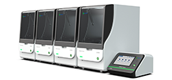
![AmMag™ Quatro Automated Plasmid Purification]() AmMag™ Quatro automated plasmid purification
AmMag™ Quatro automated plasmid purification
-
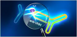
![Anti-Camelid VHH]() MonoRab™ Anti-VHH Antibodies
MonoRab™ Anti-VHH Antibodies
-
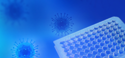
![ELISA Kits]() ELISA Kits
ELISA Kits
-
![Precast Gels]() SurePAGE™ Precast Gels
SurePAGE™ Precast Gels
-
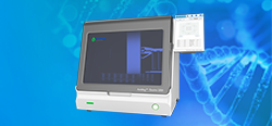
![Quatro ProAb Automated Protein and Antibody Purification System]() AmMag™ Quatro ProAb Automated Protein and Antibody Purification
System
AmMag™ Quatro ProAb Automated Protein and Antibody Purification
System
-
![TurboCHO™ Protein Expression Kit | Fast CHO Expression in 3–7 Days | GenScript]()
![Target Proteins]() Target Proteins
Target Proteins
-
![AmMag™ Quatro Automated Plasmid Purification]() AmMag™ Quatro automated plasmid purification
AmMag™ Quatro automated plasmid purification
-
![Stable Cell Lines]() Stable Cell Lines
Stable Cell Lines
-
![Cell Isolation and Activation]() Cell Isolation and Activation
Cell Isolation and Activation
-
![Quick
Order]() Quick Order
Quick Order
-
![Quick
Order]() Quick Order
Quick Order
-
- APPLICATIONS
- RESOURCES
- ABOUT US